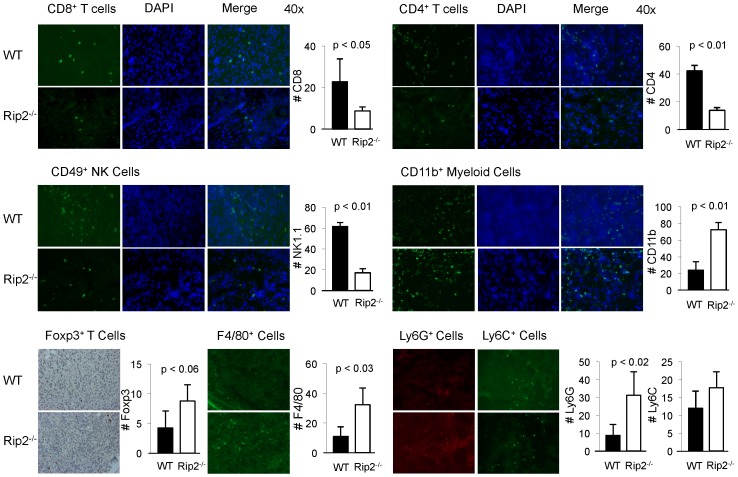
Figure 2. Loss of Rip2 alters composition of tumor infiltrating cells from Rip2-deficient mice.
Infiltration of CD8, CD4, NK, CD11b, F4/80, Ly6G, and Ly6C expressing cells as indicated, were examined by immunofluorescence, and Foxp3 by immunohistochemistry in bladder tumors from Rip2+/+ and Rip2−/− mice intravesically implanted with MB49 cells and sacrificed at 12 days. Left panels show specific antibody staining, middle panel shows DAPI staining, right panel shows merging of antibody and DAPI stains. Representative examples from each group of four to six mice in 4 independent experiments are shown at x40 magnification. Bar graphs enumerate mean number of cells per x40 field of 3 representative sections from groups of four mice; bars, SD. All p values were determined by two-tailed Student's t test, with statistically significance defined as p<0.05.

