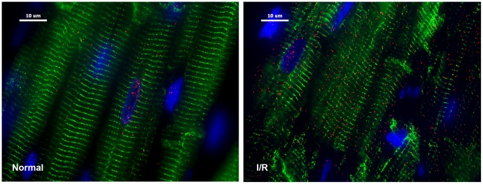
Figure 7. Chymase inside dog cardiomyocytes during ischemia/reperfusion (I/R).
Adult dogs with 60 min of LAD occlusion and 100 min of reperfusion (right panel) and normal control (left panel). I/R LV demonstrates marked increase in chymase (red) with areas of breakdown of desmin (green, right) vs. normal control tissue (left). Blue: DAPI.

