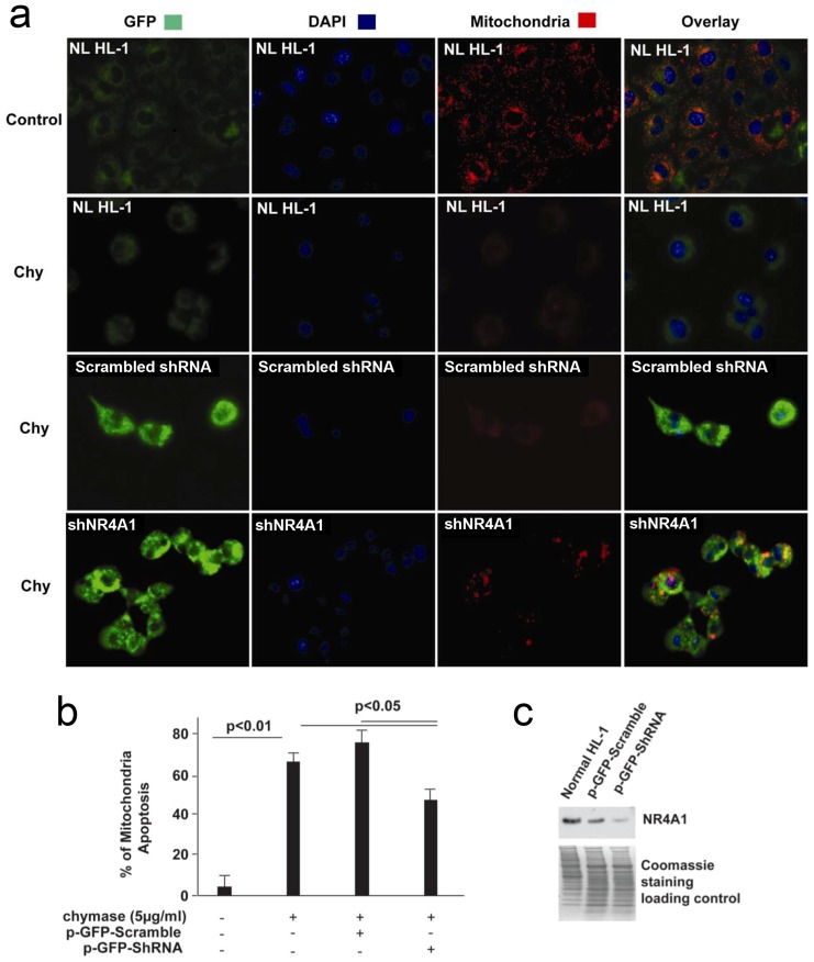
Figure 9. Knockdown of NR4A1 significantly attenuates chymase-induced mitochondria (Mt) apoptosis.
(a) Immunofluorescence pattern of HL-1 cells transfected with p-GFP-shNR4A1 and scrambled p-GFP-shRNA for 5 days, loaded with dye (MitoCapture) and then treated with 5 µg/ml chymase (chy) for 2 h. Chymase treatment induces Mt apoptosis that is detected by loss of an electrochemical gradient and the inability to take up the MitoCapture dye. Normal Mt; bright red; Apoptotic Mt: little or no red staining. Bright green is the green fluorescent protein (GFP) and indicates that the cells were transfected with either p-GFP-shNR4A1 or scrambled p-GFP-shRNA. Cells without transfection and chymase treatment are used as the control. (b) Percentage of Mt apoptosis in chymase treated HL-1 cells, HL-1 cells transfected with p-GFP-shNR4A1 and scrambled p-GFP-shRNA. (c) NR4A1 protein expression in normal HL-1 cells, HL-1 cells transfected with p-GFP-shNR4A1 and scrambled p-GFP-shRNA. NL: HL-1 cells without transfection.

