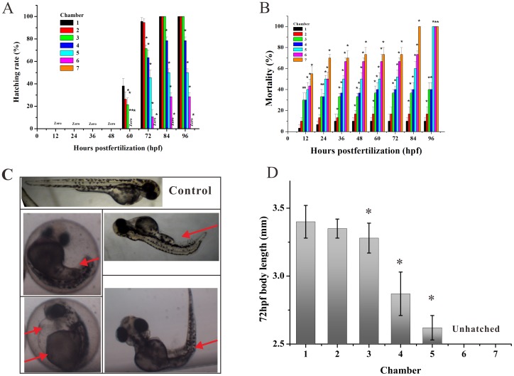
Figure 4. Apl induced abnormal morphology, developmental retardation, and mortality of zebrafish embryos.
(A) Hatched rate and (B) mortality of zebrafish embryos exposed to gradient Apl every 12 hpf in the microfluidic chip for 96 h. (C) Typical morphological abnormalities of embryos exposed to Apl. Red arrows indicate tail malformation, delayed yolk absorption, pericardial edema, and bent trunk, respectively (from upper left-right to bottom left-right). (D) Mean body length of hatched embryos treated with Apl in the chip compared with the controls (C1) at 72 hpf (n = 6), there are no date at C6 and C7 for the fish are not hatched. The asterisks indicate significant differences from control group (chamber 1) * at p<0.05.

