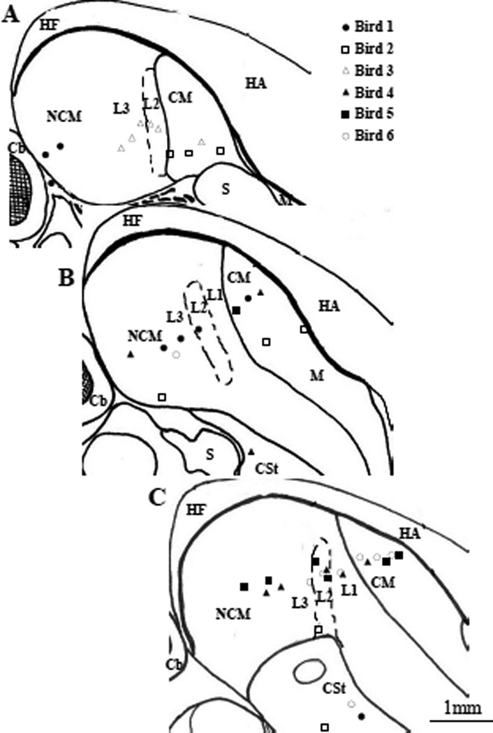
Figure 2.
Approximate locations of all electrode locations from which auditory responses were recorded. Drawings represent sagittal sections at (A) ~0.4 µm, (B) ~0.6 µm; and (C) ~0.8 µm from the midline. Electrode locations from different birds are indicated with different symbols. The rostral end of the brain is to the right, and dorsal part is at the top of the figure. Abbreviations are as in Fig. 1, except men-meninges; S-Septum.

