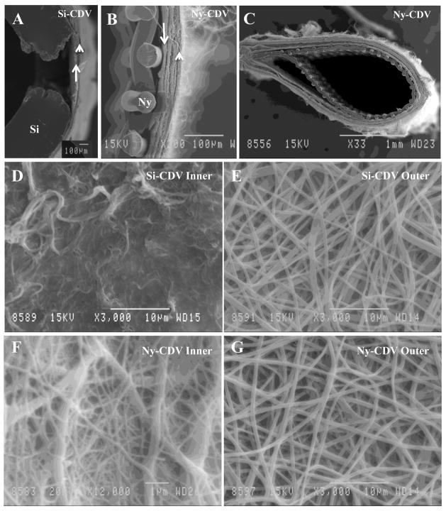
Figure 2.
Scanning Electron Microscopic Images of the Cell Delivery Vehicles A) The wall of an Si-CDV device illustrating one of the windows created in the silicone (Si) tubing using a thermal probe. The inner lining of the electrospun material is identified with an arrow and the outer layer is identified using an arrowhead. B) High magnification view of the wall of a Ny-CDV showing the inner supporting nylon mesh fabric and the two electrospun layers. The inner layer is identified by an arrow and one of the outer layers identified by an arrowhead. C) Lower magnification SEM illustrating a cross section through an Ny-CDV device D) Higher magnification SEM illustrating the inner surface of an SI-CDV device. E) Outer layer of the Si-CDV device illustrating the electrospun nylon fibers. F) Inner layer of Ny-CDV device illustrating the electrospun fibers. G) Outer layer of Ny-CDV illustrating larger fibrils and pores similar in size and density to the Si-CDV.

