
Figure 4.
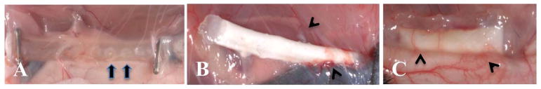
In vivo characterization of CDVs. A) Si-CDV showing ends sealed with hemoclips and loose tissue infiltration and vascularization. The windows in the silicone tubing are evident (arrows) B) Ny-CDV with cells at the time of explant showing focal areas of higher vascularity (arrowheads). C) LinBit insulin pellets within a dual electrospun membrane Ny-CDV implanted subcutaneously. The extensive peri-implant angiogenic response is evident (arrowheads).