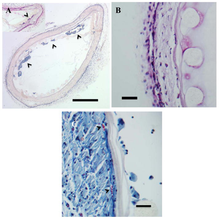
Figure 5.
In vivo characterization of CDVs. Hematoxylin and eosin stained sections of Ny-CDV devices implanted in a subcutaneous position in the mouse.. A) Ny-CDVs showing retention of delivered HaCaT (arrowheads) within the Ny-CDV. No breach of the inner cell isolation layer is apparent and no evidence of HaCat cells outside the device was found. Note that the inner nylon sheet is for structural support only and the pores are large enough to allow cells to move across. Inset shows one of the devices with cells at the inner electrospun surface. (Bar = 500 μm) B) Ny-CDV illustrating the components of the CDV: 1- Nylon mesh with the cylindrical fibers, 2- Inner low porosity electrospun layer, 3- Outer high porosity and tissue integration layer. Cells are evident penetrating through the outer porous electrospun layer but do not penetrate the low porosity layer. (Bar = 50 μm). C). Masson’s trichrome stained section of a Ny-CDV illustrating the neovascularization of the porous electrospun layer. Arrowheads identify two vascular profiles at the interface between the high porosity and low porosity electrospun coating. (Bar = 50 μm).

