
Figure 6.
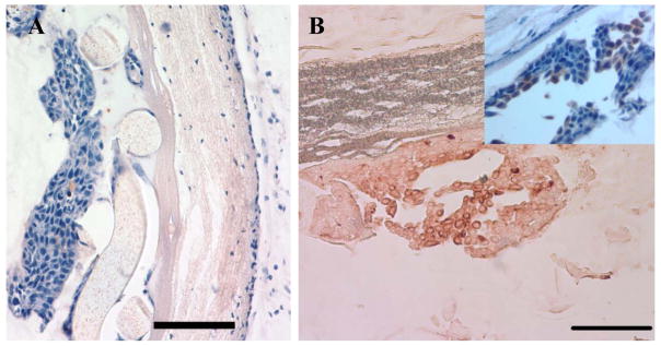
Cellular isolation in the CDV in vivo. A) Light photomicrograph of a hematoxylin and eosin stained section of a Ny-CDV illustrating cellular isolation of human HaCaT epithelial cells separated from the host tissue by the intact inner low porosity membrane. (Left to Right: CDV lumen to host tissue) (Bar = 100 μm) B) Cytokeratin 18 expressing HaCaT cells exhibit a brown reaction product and are localized within CDV lumen (Scale 100μm). Inset: HaCaT cells counterstained with hematoxylin to illustrate the localization of the HaCaT cells within the lumen of the device and the viability of the cells.. (Bar = 100 μm)