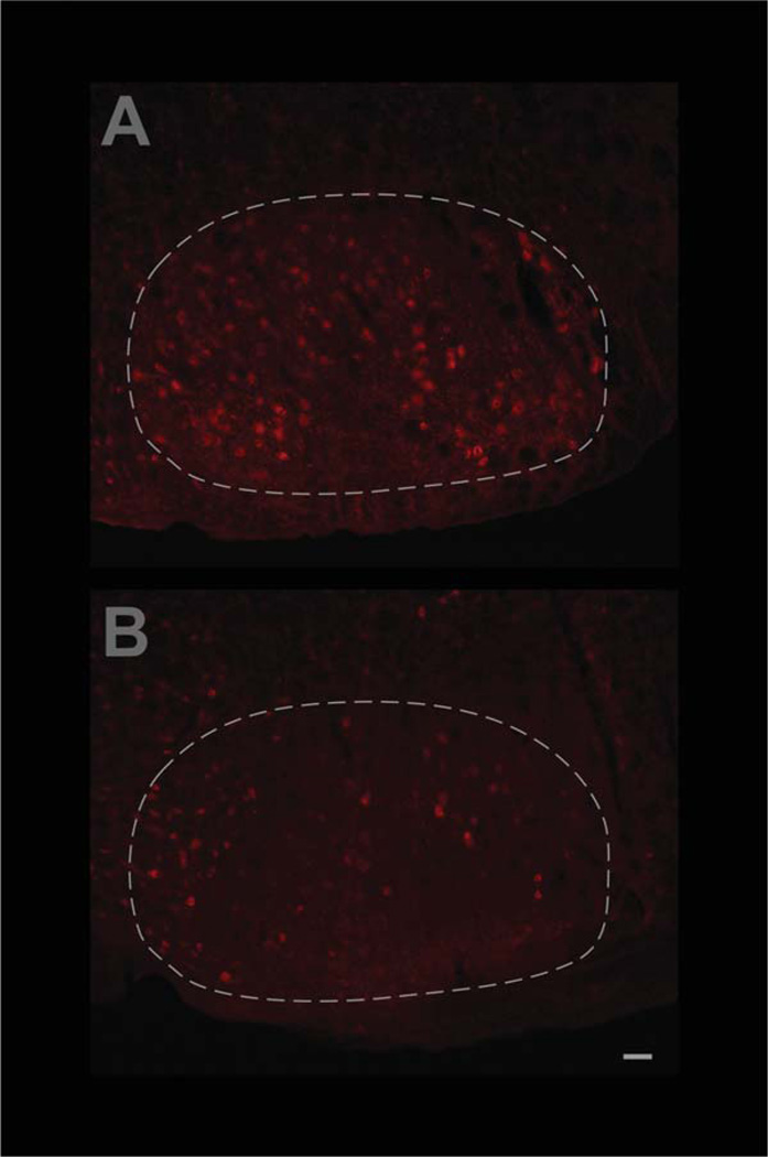
Figure 2.
Confirmation that adult-onset CNTFRα gene excision in facial motor neurons leads to the expected loss of CNTFRα. CNTFRα-floxed mice and littermate controls, all containing the inducible Cre gene construct, CreER, were simultaneously injected with tamoxifen at 3–4 months of age and their facial motor nuclei were examined by CNTFRα immunohistochemistry 3 months later. Examples of sections from control (A) and CNTFRα-floxed (B) mice are presented. Quantification of all the data by investigators blind to genotype indicated that the floxed mice displayed only 55.9 ± 25.8% as many CNTFRα-labeled facial motor neurons as the littermate controls processed in parallel (P < 0.05; t = 3.41; n = 4 pairs). Images in (A,B) were identically captured and adjusted. The broken white line delineates the approximate borders of the facial motor nucleus. Scale bar = 50 µm.

