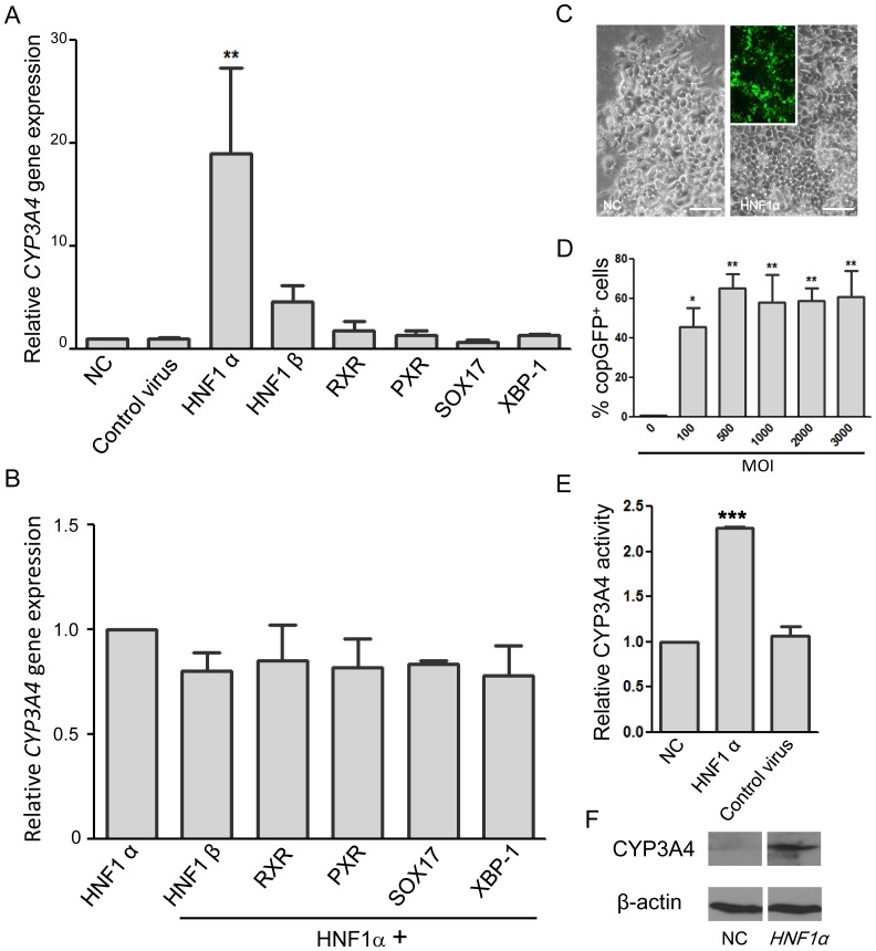
Figure 1. Selection of HNF1α as the optimal regulator for induction of CYP3A4 expression in Hep G2 cells.
Cells were infected by control or indicated recombinant lentiviruses and cultured for 7 days before determination of the CYP3A4 expression and activity. (A) CYP3A4 mRNA expression in normal control (NC) Hep G2 cells and cells infected (at MOI = 100) with control virus or recombinant lentiviruses respectively expressing HNF1α, HNF1β, RXR, PXR, SOX17, or XBP-1. Optimal CYP3A4 expression occurs in cells infected with the HNF1α-expressing lentivirus. (B) Co-infection of Hep G2 cells with the HNF1α lentivirus and either HNF1β, RXR, PXR, SOX17, or XBP-1 did not increase CYP3A4 mRNA expression levels. (C) Left panel: A representative bright field image of control HepG2 cells. Right panel: A representative bright field image and fluorescent image of HepG2 cells infected at MOI = 3,000 by lentiviruses containing HNF1α, showing the reporter copGFP fluorescence. Scale bar = 100 µm. (D) Flow cytometry analysis showing percentage of copGFP+ Hep G2 cells 7 days post-infection with HNF1α-expressing lentivirus at various MOI. (E) Increased CYP3A4 enzyme activity and (F) protein levels in Hep G2 cells infected with the HNF1α-expressing lentivirus (MOI = 100) compared with normal control cells (NC) or cells infected with control virus. **P<0.01, ***P<0.001 compared with NC cells.

