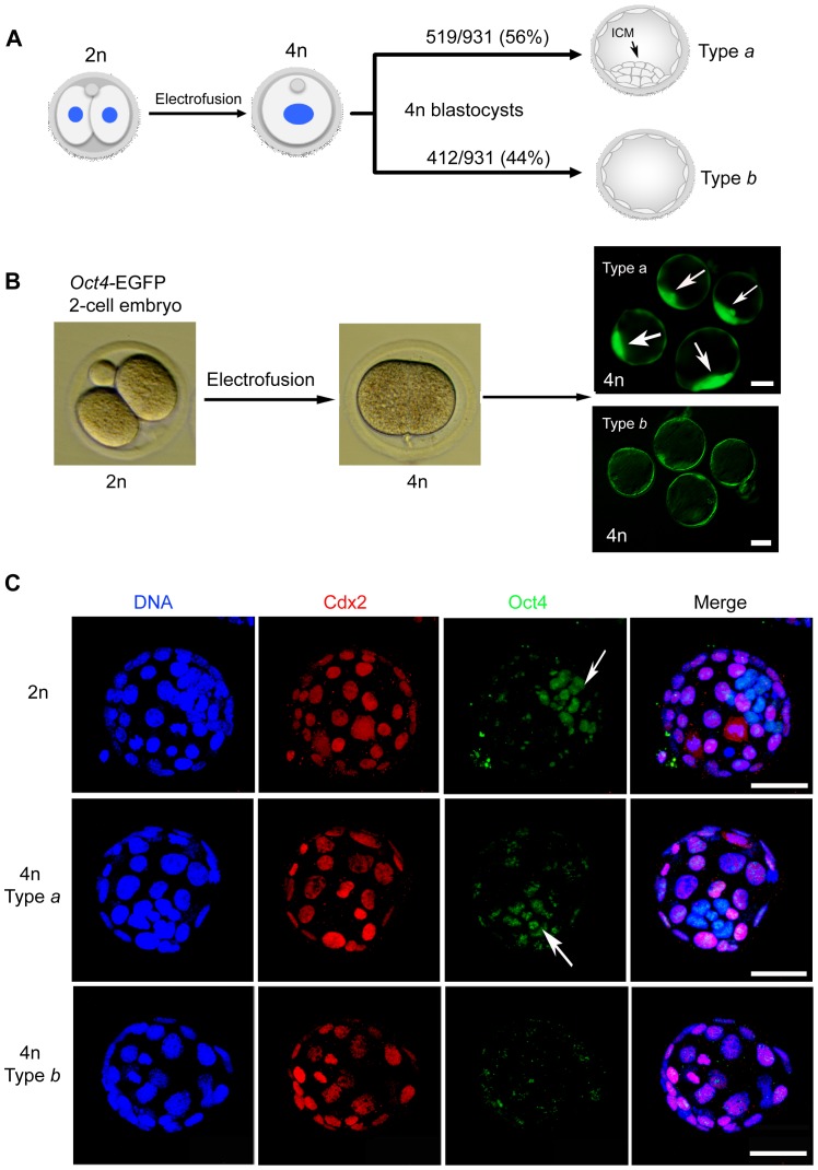
Figure 1. Mouse tetraploid blastocysts are grouped into two types.
(A) Schematic illustration of tetraploid embryo generation. The two blastomeres of 2-cell stage diploid (2n) embryos are electrofused into one large blastomere thus doubling the DNA content to tetraploid (4n) in the embryos. The resulting 4n embryos can normally develop to blastocysts and are classified into two groups by the presence (type a) or absence (type b) of an ICM. (B) The ICM in 4n blastocysts of Oct4-EGFP embryos can be visualized by expression of EGFP in the resulting 4n blastocysts, and are classified into type a or type b under the fluorescence microscope. (C) Confocal images of diploid and tetraploid type a and tetraploid type b blastocysts, images are full projections of 20 optical sections. Embryos were stained with antibodies of CDX2 (staining the trophoblast) and OCT4 (staining the ICM). Both the diploid and tetraploid type a blastocysts showed ICM in the embryos, whereas the tetraploid type b blastocysts lacked an ICM. Arrow indicates the ICM. Scale bar: 50 µm.

