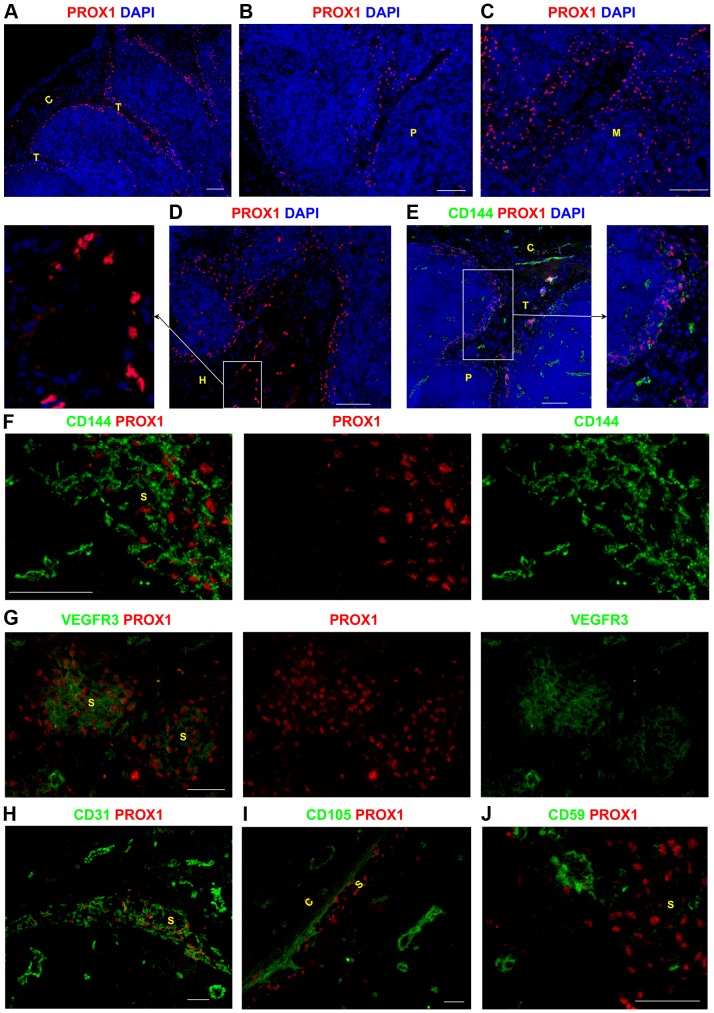
Figure 1. Identifying lymphatic sinuses in the human lymph node using specific markers of lymphatic endothelial cells.
Frozen LN sections were probed with an antibody to detect the transcription factor PROX1 to map the lymphatic sinuses in the human LN. The subcapsular and trabecular sinuses were PROX1+ (A). PROX1+ sinuses were also found in the paracortex (B), the medulla (C), and near the hilum (D). CD144 and VEGFR3 were expressed by lymphatic structures however they were not exclusive LEC markers as they were also expressed by BECs (E–G). Lymphatic sinuses in the medulla expressed varying levels of VEGFR3 (G). Similarly CD31 was also expressed by LECs and BECs throughout the LN (H). LECs lacked expression of CD105 (I) and CD59 (J), whilst these markers were clearly expressed by blood vessels. Blue represents DAPI staining of cell nuclei (A–E). C,capsule; T, trabecula; P, paracortex; M, medulla; H, hilum; S, sinus. Scale bars represent 100 µm (A–E) and 50 µm (F–J).

