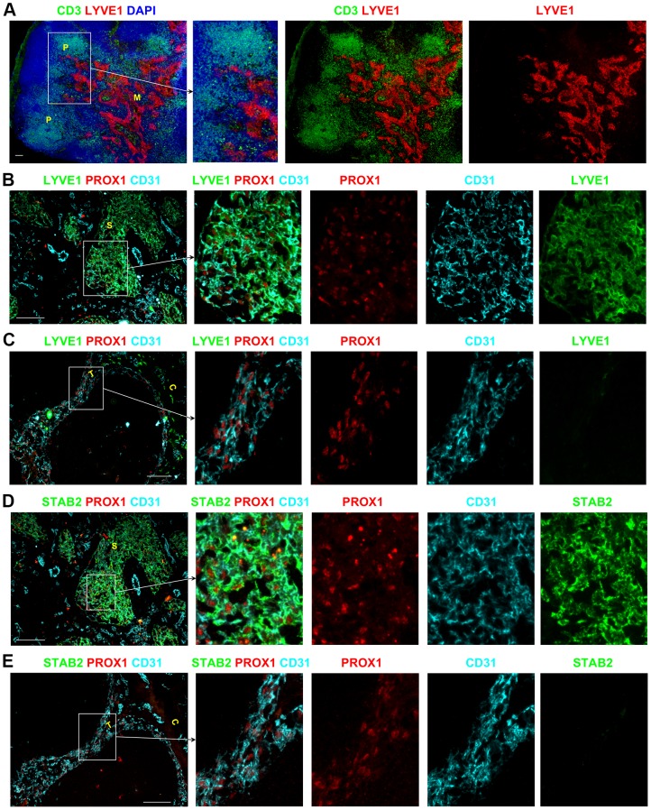
Figure 2. Heterogeneous expression of LYVE1 and STAB2 by endothelial cells in lymphatic sinuses of human LNs.
Low magnification images demonstrated that LYVE1 expression is restricted to the paracortical and medullary sinuses whilst the other sinuses in the superficial areas lack expression of this marker (A). PROX1+CD31+ LECs of the paracortical and medullary sinuses express LYVE1 (B) and STAB2 (D), whereas the LECs in the subcapsular and trabecular sinuses are negative for LYVE1 (C) and STAB2 (E). Blue represents DAPI staining of cell nuclei (A). P, paracortex; M, medulla; C, capsule; T, trabecula; S, sinus. All scale bars represent 100 µm.

