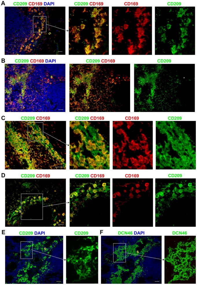
Figure 5. CD209 expression in lymphatic sinuses.
CD169+ APCs also expressed CD209 (A–D). Rare CD169+ CD209− APCs were detected in one of the LNs assessed (B: middle panel marked with asterisk). CD209+CD169− cells in the paracortical and medullary sinuses are likely to represent LECs (C–D). Serial LN sections stained with anti-CD209 (E) and DCN46 (F) showed that far fewer cells were stained by anti-CD209 antibody than by DCN46. Blue represents DAPI staining of cell nuclei (A–B, E–F). C,capsule; S, sinus. All scale bars represent 50 µm.

