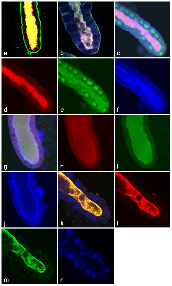
Figure 7. Evidence for the graded temporal release of different proteins by apocrine secretion.
(a) = At+8.5 hr APF, the ribosomal protein Rp40 (blue) is completely released into lumen, the cortical membrane component α-spectrin (green) was removed from the lateral and apical surfaces but remained at the basal surface, and the nuclear receptor Usp (red) is about half-released into the lumen. (b) At +9 hr APF, both the ribosomal protein Rp21 (green) as well as the ecdysone-inducible Ets-like E74 transcription factor (red) are present only in the lumen, whereas there remains significant F-actin (blue) signal on the cortical membranes. (c) At the same time (+9 hr APF), the ecdysone-regulated transcription factor and nuclear tumor suppressor are secreted differently: while Kr-h (red (d)) is completely extruded into the lumen, p53 (green (e)) only starts to be released and the majority of its signal is still detected in nuclei. Although filamentous actin (blue (f)) already is being secreted into the lumen, there is detectable signal still visible on cell membranes. (g) During +9 to +10 hr APF, the ecdysone-regulated transcription factor BR-C (green (h)) is completely released into the lumen, whereas lamin C (red), a component of the nuclear envelope, is only partially released and can be still detected on the nuclear membrane (i). Although filamentous actin (blue) is already within the lumen, significant amounts of it still line the cortical cytoskeleton, mainly at the apical membrane (j). (k) At the end of +10 hr APF both, Rab11 (green (l)), a member of the GTPase family of membrane proteins as well as the tumor suppressor transcription factor p53 (red (m)) have been completely secreted into the lumen. Hoechst 33258 was used to detect nuclear DNA (blue (n)) which stays in nuclei. All confocal images 400×.

