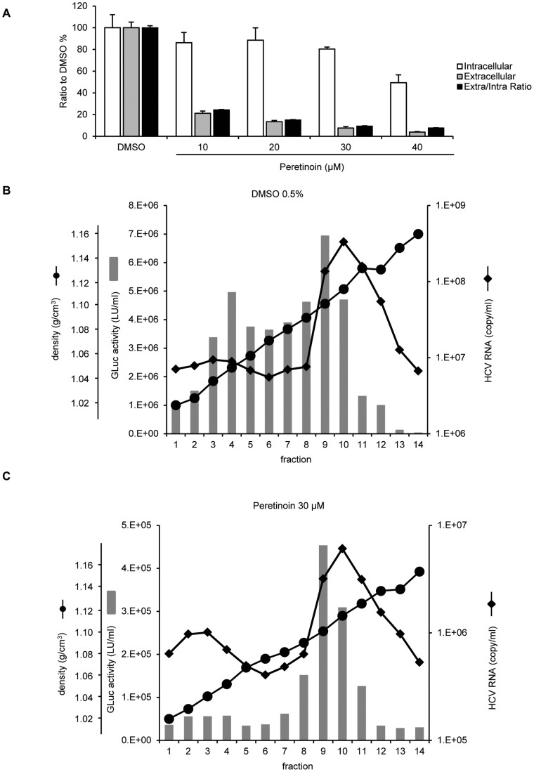
Figure 6. Impact of Peretinoin on infectious virus production.
(A) FT3-7 cells were transfected with HJ3-5/GLuc2A RNA, and 7 days later, 0.5% DMSO, or 10–40 μM Peretinoin, were added. At 72 h later, extra- and intra-cellular viruses were collected and used to infect naïve Huh-7.5 cells. Replication capacity was also determined by measuring secreted GLuc activity. At 48 h after infection, we determined the amount of infectious virus from extra- and intra-cellular media by using GLuc activity as an indicator of the amount of infectious virus. Intra- and extra-cellular infectivity was normalised to replication capacity at infection, and these were then normalised to those of DMSO-treated cells, which were set to 100%. The ratio of extracellular infectious virus to intracellular virus was calculated at the indicated conditions, and it was then normalised to DMSO-treated cells, which were set to 100%. Data show the mean ratio to that of DMSO-treated cells ± SD from 3 independent experiments. (B, C) FT3-7 cells were transfected with HJ3-5/GLuc2A RNA, and 7 days later, 0.5% DMSO or 30 μM Peretinoin were added, and then 72 h later, the medium was collected and subjected to equilibrium ultracentrifugation. Fourteen fractions were taken and analysed for density (circles), HCV RNA levels (diamonds), and infectious virus titres determined by GLuc activity (grey bars). (B) shows the results from DMSO-treated cells, whilst (C) shows those for Peretinoin-treated cells.

