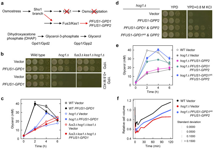
Figure 2. Synthetic osmoadaptation in hog1Δ cells.
(a) Experimental design for synthetic osmoadaptation in hog1Δ using crosstalk and the glycerol biosynthesis pathway. Expression of Hog1-dependent osmostress-induced genes (GPD1 or/and GPP2) was rewired under the control of a Fus3/Kss1 dependent FUS1 promoter. (b) Osmotic induction of GPD1 expression via crosstalk partially suppresses osmosensitivity of hog1Δ in a Fus3/Kss1-dependent manner. Cells of the indicated strains carrying YIp352 or YIp352-PFUS1-GPD1 were grown on YPD plates with or without 0.8 M KCl for 1–2 days at 30°C. (c) Intracellular glycerol accumulation correlates with the cell growth shown in (b). Cells were grown to mid-log phase, subjected to osmotic stress (0.8 M KCl), and intracellular glycerol was monitored at the indicated time points. Values represent the mean and standard deviation of three replicas. (d) Osmotic induction of GPD1 and GPP2 together via crosstalk strongly suppresses osmosensitivity of hog1Δ. Cells of the hog1Δ strains carrying YIp352-PFUS1-GPP2 and/or YIplac128-PFUS1-GPD1 (or GPD14A) were grown as in (b). Cell growth shown in (d) correlates well with the ability of those cells to accumulate glycerol (e) and recover cell volume (f) after osmotic treatment. For cell volume data, line thickness indicates standard deviation of data obtained with approximately 30 cells. See Methods for the details of glycerol assay and cell volume measurement.

