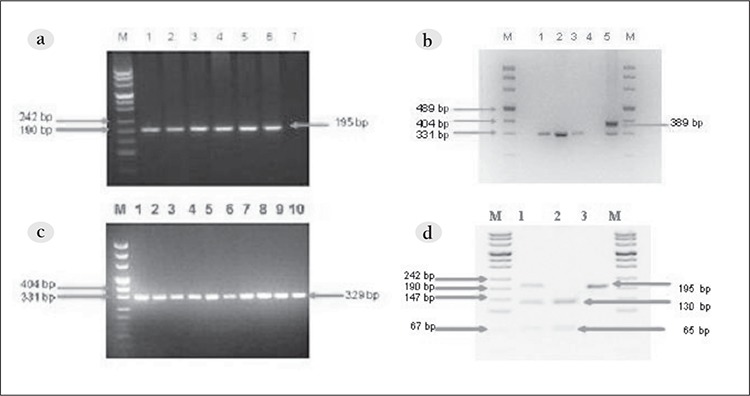
Figure 1. a. PCR amplification of the FLT3 -ITD region (lane M: size marker; lanes 1-10: normal samples). b. PCR amplification of the FLT3 -ITD region (lane M: size marker; lanes 1-3: normal samples; lane 4: negative control; lane 5: FLT3 /ITD-positive case).c. PCR amplification of FLT3 -D835 (lane M: size marker; lanes 1-6: normal samples; lane 7: negative control). d. D835 mutation detection (lane M: size marker; lane 1; FLT3 -D835-positive case; lane 2: wild type; lane 3: EcoRV undigested sample).

