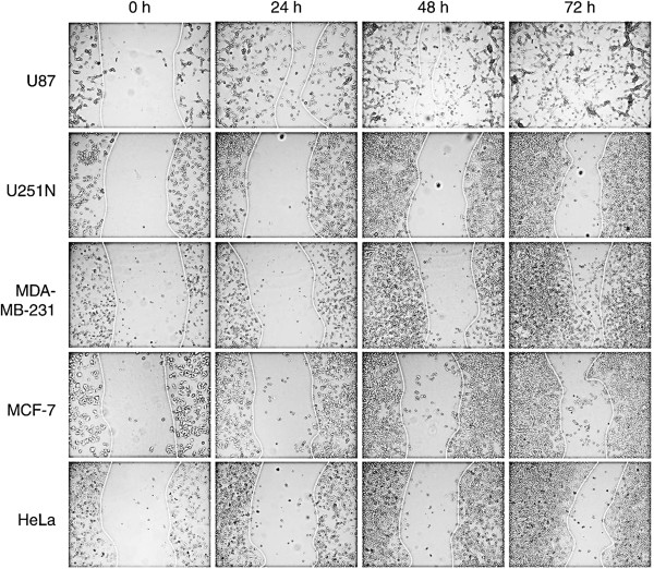
Figure 2.
Cancer cell migration using ring cell migration assay. Cells (250,000 to 300,000) were seeded in 6-well plates with cloning rings. After removal of rings, photographs were taken of the gaps observed under microscope at different time points, i.e. 0 h, 24 h, 48 h and 72 h. The dotted lines delineate the migrating edges of cells.

