Figure 1.
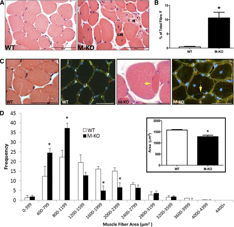
Myopathic features of β1β2M-KO muscles. A) H&E images of WT and β1β2M-KO (M-KO) skeletal muscles. Note the numerous central nucleated (CN) fibers, diverse fiber shapes, and presence of necrotic (N) fibers with excess endomesial tissue. B) When quantified, there was a significant increase in the total number of fibers containing a central nuclei in the β1β2M-KO tibialis anterior muscles. C) Another general characteristic of muscle myopathy is the presence of split fibers. In this H&E image, you can note the presence of a split fiber in the β1β2M-KO muscle, while this feature was absent in WT muscle. This was confirmed with an immunofluorescent stain for dystrophin (green; sarcolemma), laminin (red; basal lamina), and DAPI (blue; nuclei). In a split fiber, the basal lamina will surround the entire (both) fibers, while the dystrophin will surround each fiber separately. Yellow arrow highlights the presence of dystrophin with no overlying basal lamina. Split fibers were consistently observed in the β1β2M-KO muscle cross sections but were not observed in WT muscles. D) Myofiber cross-sectional area of WT and β1β2M-KO TA muscles were measured and binned according to size. Note the significant increase in smaller fibers and significant decrease in larger fibers in β1β2M-KO muscles. On average, the mean cross-sectional area of β1β2M-KO muscle fibers was significantly reduced compared to WT (inset). Data are expressed as means ± se; n = 3 or 4. Scale bars = 50 μm (A); 60 μm (C). *P < 0.05 vs. WT.

