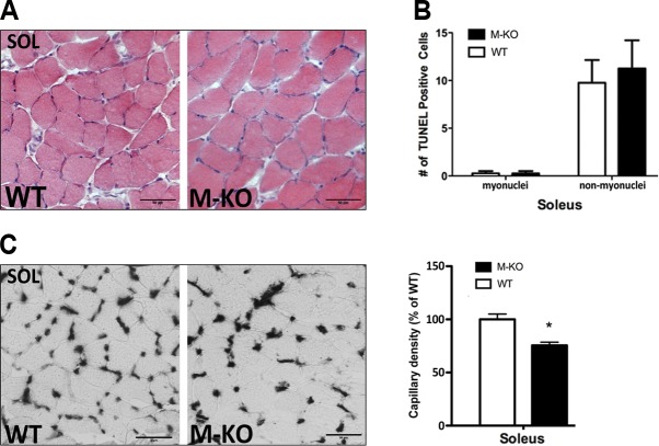
Figure 5.
β1β2M-KO soleus muscles display no myopathy and only modest reductions in capillary density. A) There is an absence of myopathic traits in soleus (SOL) muscles of β1β2M-KO (M-KO) mice, as can be seen in these H&E stains. B) Quantification of the TUNEL-positive nuclei within the soleus muscle demonstrated no difference in either muscle or nonmuscle nuclei that are TUNEL-positive. C) Representative alkaline phosphatase stains (left panel) and quantification of capillary density in soleus muscles of WT and β1β2M-KO mice. Reduction in capillary density is much less (only ∼20%) in soleus compared to TA muscles of β1β2M-KO mice. Data are expressed as means ± se; n = 3–4. Scale bars = 50 μm. *P < 0.05.

