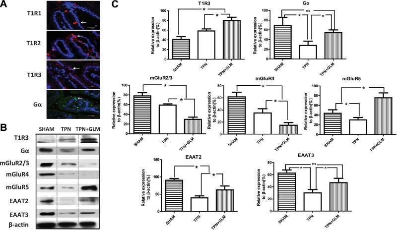
Figure 1.
Influence of TPN administration and GLM supplementation on the amount of luminal sensing molecules of isolated IECs at the protein level. A) IF staining was performed on jejunal mucosa from TPN mice with anti-T1Rs and anti-Gα Abs and counterstained with DAPI nuclear stain. Note the similar localization of 3 T1Rs and Gα proteins in the cytoplasm of the endocrine-like solitary cells of the jejunal villi. B, C) Protein expression of T1R3, mGluRs, transporters, and Gα in sham-treatment mice compared with TPN and TPN + GLM (75 mM) mice. B) Representative immunoblots of isolated small-bowel IECs from each group. C) Mean protein level for each group. For representative images and immunoblots, a minimum of 3 experiments and images were used for each selection. Scale bars = 50 μm. Values are means ± sd (error bars). ns, not significantly different. *P < 0.05.

