Figure 5.
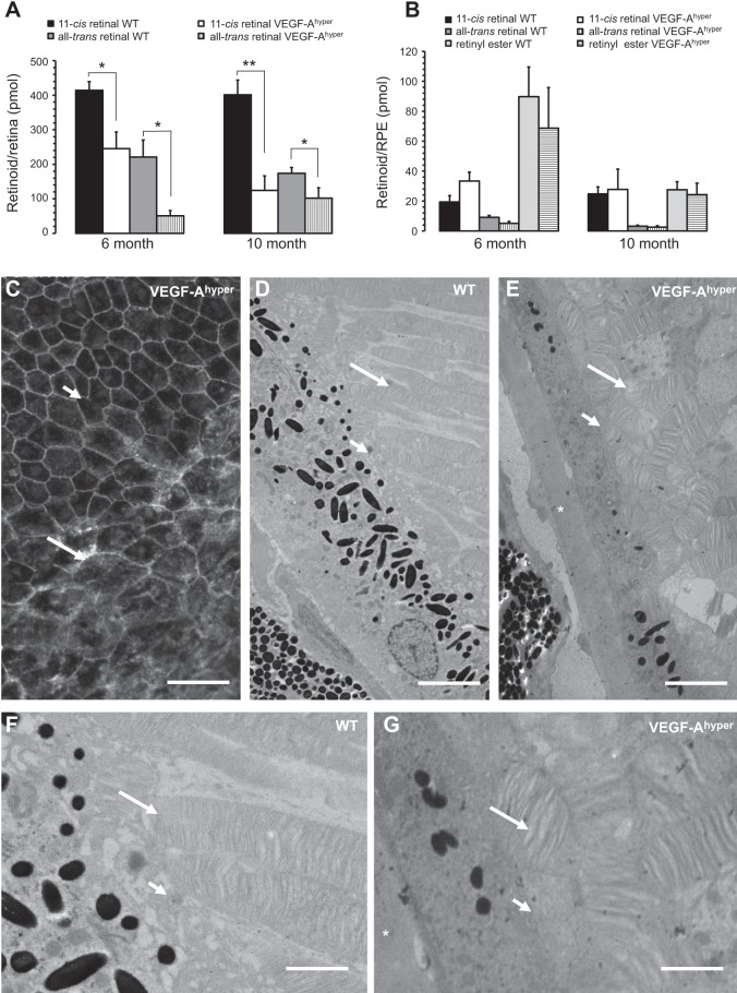
Retinoid profiling reveals a defect in the visual cycle with reduction of 11-cis and all-trans retinal levels in the retinas of VEGF-Ahyper mice. A) Retinas of VEGF-Ahyper mice show a reduction of 11-cis and all-trans retinal, which is more pronounced in 10-mo-old mutant mice compared to 6-mo-old mice. Bars represent means ± sd. *P < 0.05, **P < 0.01. B) Retinyl esters in the RPE (RPE/choroid) do not show a significant difference between VEGF-Ahyper mice and control mice. Bars represent means ± sd. C) VEGF-Ahyper mice with these visual cycle defects show an RPE barrier breakdown, demonstrated here by cytoplasmic accumulation of β-catenin staining and loss of typical RPE cell honeycomb morphology (long arrow). Adjacent nonlesional RPE cells (short arrow) maintain normal RPE cell morphology in young (2-mo-old) VEGF-Ahyper mice. Adapted from ref. 4. D–G). Defects in visual cycle correlate with abnormalities in the interdigitation of apical RPE cell membranes with photoreceptor outer segments in aged (15-mo-old) VEGF-Ahyper mice, shown by electron microscopy. Photoreceptor outer segments in mutant mice (E, G) are highly disorganized and shortened, compared to WT mice (D, F). Long arrows show photoreceptor outer segments. Short arrows show apical RPE cell membranes. Asterisks indicate electron-dense basal laminar sub-RPE deposits. Panels F and G are magnifications of images in D and E, respectively. Scale bars = 75 μm (C); 3 μm (D, E); 1.5 μm (F, G).

