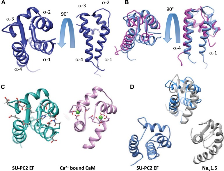
Figure 3.
A) Structure of Ca2+-bound suPC2-EF and comparison to other EF structures. Backbone ribbon for the top model (taken from the top 20 conformers). B) Comparison of suPC2-EF (blue) and hPC2-EF (magenta; PDB ID: 2KQ6). C) Side-by-side comparison of suPC2 and CaM (PDB ID: 1CLL) with the residues that coordinate Ca2+ represented with Corey-Pauling-Koltun (CPK) stick models. CaM shows the coordinating residues, with green Ca2+ spheres. D) Overlap comparison and side-by-side comparison of the ribbon structures of suPC2-EF (blue) and NaV1.2 EF (gray; PDB ID: 4DCK).

