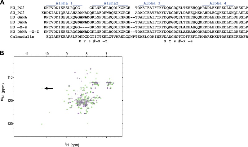
Figure 4.
Insertion of DANAD into hPC2-EF results in two Ca2+ binding sites. A) Constructs and the mutants created for this study. B) Overlaid 1H-15N HSQC NMR spectra of hPC2-EF (violet contours) and DANA hPC2-EF (green contours) at 600-MHz proton frequency. Proteins were in 25 mM TRIS and 150 mM KCl buffer (pH 7.4). Arrow indicates amide protons involved in hydrogen bonding with the side-chain carboxyl oxygen atom of the corresponding aspartate residue in the DANA construct, indicating a second Ca2+ binding site.

