Figure 1.
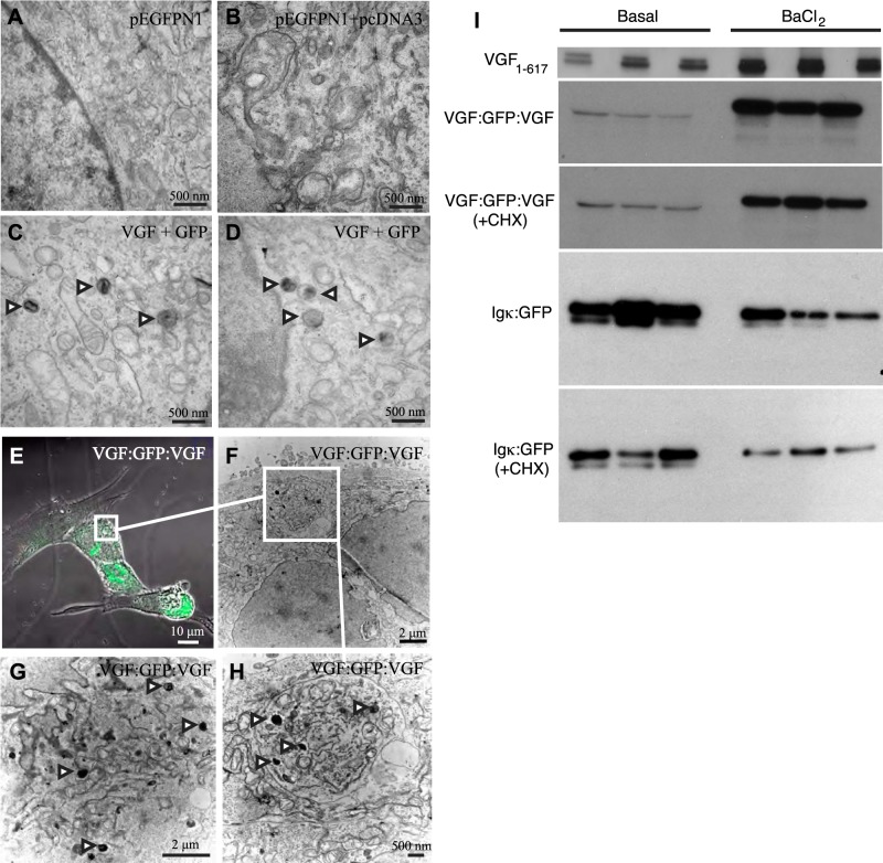
Expression of exogenous VGF in nonendocrine NIH 3T3 cells results in the formation of LDCV-like vesicles, visualized by CLEM, and regulated secretion of VGF. A–D) NIH 3T3 cells were cotransfected with plasmids encoding GFP and either full-length rat VGF or empty vector, and identified GFP-positive cells were processed for transmission EM as detailed in Materials and Methods. Representative transmission electron micrographs of 3T3 cells transfected with pEGFPN1 alone (A), and those cotransfected with pEGFPN1 and empty pcDNA3 vector (B), or pEGFPN1 and rat VGF1–617 expression plasmids (C, D), are shown. Arrowheads indicate LDCV-like structures. E–H) NIH 3T3 cells were similarly transfected with plasmid encoding VGF1–65:GFP:VGF452–617 (VGF:GFP:VGF), and LDCV-like structures were visualized in GFP-positive, green cells (E) by CLEM (F–H). Arrowheads indicate LDCV-like structures. I) To measure secretion, NIH 3T3 cells were transfected with expression plasmids encoding rat VGF1–617, VGF1–65:GFP:VGF452–617 (VGF:GFP:VGF), or constitutively secreted Igκ:GFP (26). After 48–72 h, cells were rinsed, incubated for 30 min in serum-free medium (basal release), rinsed, and incubated for 30 min in serum-free medim supplemented with 2 mM BaCl2 (stimulated release). In a subset of experiments, cells were pretreated for 2 h with CHX (1 μg/ml), which blocks constitutive but has less effect on regulated secretion, or with vehicle, and basal or stimulated release was assayed as described above, in the presence of CHX or vehicle. Secreted VGF, VGF:GFP:VGF, or Igκ:GFP proteins were quantified by Western blot analysis and ImageJ, as described in Materials and Methods. Normalized to basal release (1±0.2), VGF secretion was stimulated 2.8 ± 0.1-fold by BaCl2 (P=0.004, 2-tailed Student's t test). VGF:GFP:VGF secretion under basal conditions (1±0.3) was stimulated 18.6 ± 1.3-fold by BaCl2 (P<0.001, ANOVA with Tukey's post hoc comparison, n=3), while basal + CHX release (2±0.4) was stimulated 12.2 ± 0.7-fold in BaCl2 + CHX-treated cells (P<0.001, ANOVA with Tukey's post hoc comparison, n=3) (all values normalized to VGF:GFP:VGF secretion under basal conditions). Igκ:GFP secretion under basal conditions (1±0.12) was not stimulated by BaCl2 treatment but rather was significantly reduced (0.4±0.14; P<0.05, ANOVA with Tukey's post hoc comparison, n=3). Secretion of Igκ:GFP in the presence of CHX (0.5±0.07) was not stimulated by BaCl2 + CHX (0.2±0.03; nonsignificant compared to CHX). Igκ:GFP secretion in BaCl2 + CHX-treated cells was significantly reduced compared to basal secretion (P=0.0025) (all values normalized to Igκ:GFP secretion under basal conditions). Results are representative of ≥3 experiments.

