Figure 3.
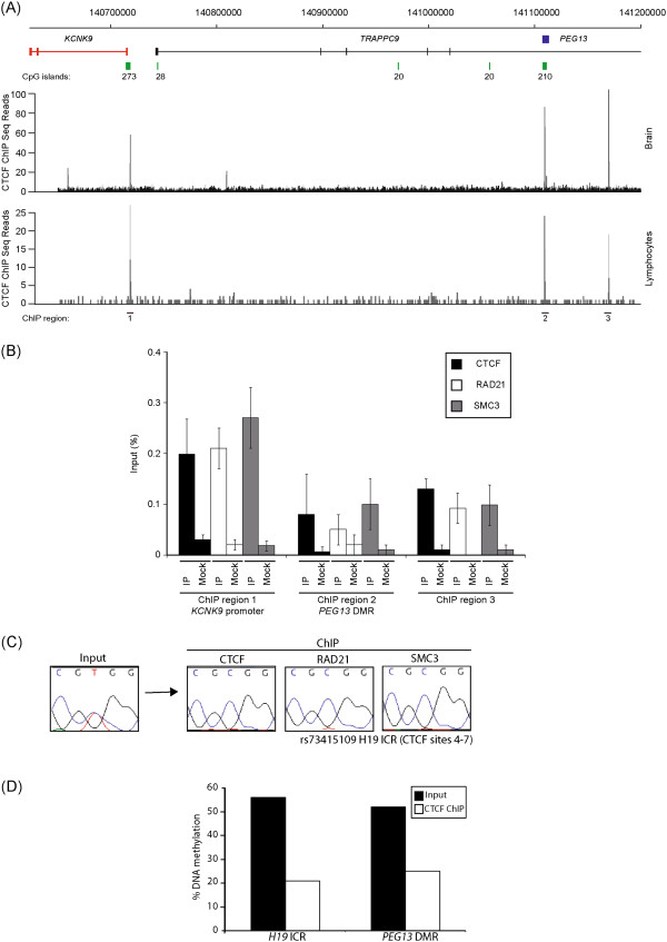
ChIP analysis for CTCF and the cohesion subunits RAD21 and SMC3. (A) ChIP-Seq data analysis in lymphocytes and cerebellum reveals the location of three ubiquitous CTCF binding sites. The positions of the ChIP PCRs are indicated. (B) qPCR performed on CTCF, RAD21 and SMC3 ChIP material in normal lymphoblastoid cells at the intervals identified by ChIP-Seq. Graphs are represented as % of precipitation relative to input chromatin (mean values ± SEM). (C) Sequence traces showing monoallelic precipitation of CTCF and cohesion subunits at the control H19-ICR. (D) The methylation levels of at the H19-ICR and PEG13-DMR in CTCF input and ChIP material as determined by bisulphite PCR followed by pyrosequencing.

