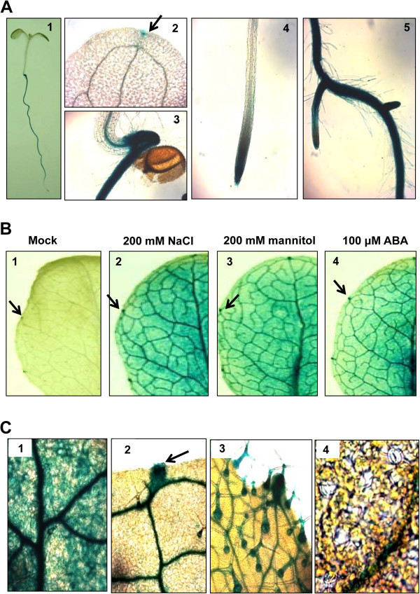
Figure 3.
Tissue-specific expression of GUS in APUM5pro-GUS transgenic plants. (A)APUM5 promoter activity was determined by GUS histochemical staining. Section 1, 4-day-old seedlings. Section 2, hydathode on the cotyledon indicated by an arrow. Section 3, shoot apical meristem region. Section 4, primary root. Section 5, lateral roots. (B) The effect of salt, osmotic, and ABA treatments on APUM5pro-GUS activity. Three-week-old seedlings were treated with 200 mM NaCl, 200 mM mannitol, or 100 μM ABA for 6 h before GUS staining. Enhanced APUM5 promoter activity on the rosette leaf following abiotic stress. (C) Enlarged image from 3B. Section 1, APUM5 promoter activity expressed in leaf vasculature and mesophyll cell regions. Section 2, hydathode and leaf vascular bundle. Section 3, trichomes on a rosette leaf. Section 4, guard cells.

