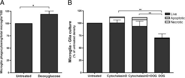
Figure 7.

Microglial phagocytosis contributes to the loss of microglia induced by deoxyglucose. (A) Pure primary microglia were treated ± deoxyglucose (DOG; 10 mM) for 21 hours, and were then incubated with 5 μm carboxylate-modified latex microspheres for 2 hours, and the number of beads phagocytosed per cell quantified. (B) The phagocytosis inhibitor cytochalasin D (1 μM) decreased the loss of microglia measured 6 hours after the addition of DOG (10 mM) in glial cultures. Data presented as mean ± standard error of the mean for ≥ 3 independent experiments. *Total microglia: *P < 0.05, **P < 0.01.
