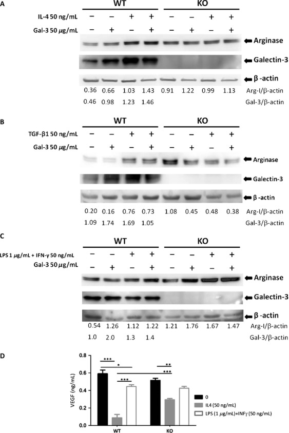
Figure 5.

(A, B and C) Western blotting WT-BMDM or KO-BMDM of total protein cell extracts without stimulation or after IL-4 (50 ng/mL), TGFβ1 (50 ng/mL) and LPS (1 μg/mL) + IFN-γ (50 ng/mL), with or without exogen galectin-3 (50 μg/mL). Each lane represents a pool from three independent assays (50 μg/lane), each one performed with cells derived from one animal. The images were representative of two independent experiments. The number above each lane represents the target/β-actin relation from densitometric analysis performed using ImageJ. (D) The ELISA evaluated VEGF secreted in medium from bone marrow-derived macrophages WT or KO cells after 24 h of culture. The experiments were conducted in triplicates comparing basal levels with M2 polarization (IL-4, 50 ng/mL) or M1 polarization stimuli (LPS, 1 μg/mL + IFN-γ, 50 ng/mL). Similar results were obtained in a second experiment, consisting of an analysis of pooled samples from three independent plates of BMDM, each one obtained from different animals. WT, wild type; KO, knockout; BMDM, bone marrow-derived macrophages; ELISA, enzyme-linked immunosorbent assay.
