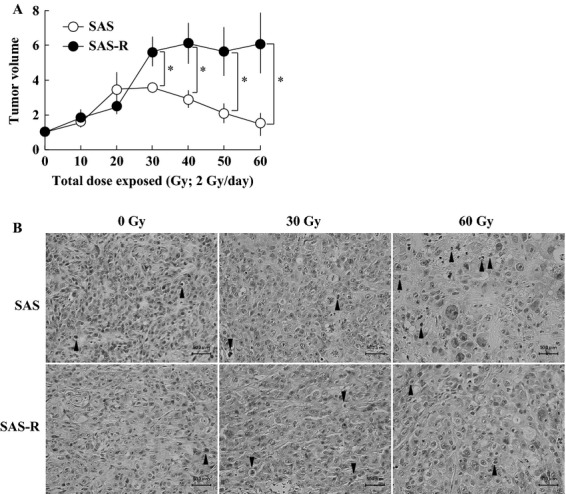
Figure 1.

(A) Establishment of a clinically relevant radioresistant tumor model. Exponentially growing SAS and SAS-R cells were subcutaneously injected into the dorsal flank of nude mice. When tumor volumes reached about 150 mm3 (day 0), mice were exposed to FR with 2 Gy/day of X-rays for 30 days. Mean ± SD of three independent mice are shown. *P < 0.01. (B) Histological analysis of SAS and SAS-R tumors exposed to 2 Gy/day of fractionated X-rays. Connective tissues were more abundant in SAS-R tumors than in SAS tumors. Arrow heads, pyknotic cells. FR, fractionated radiation.
