Figure 6.
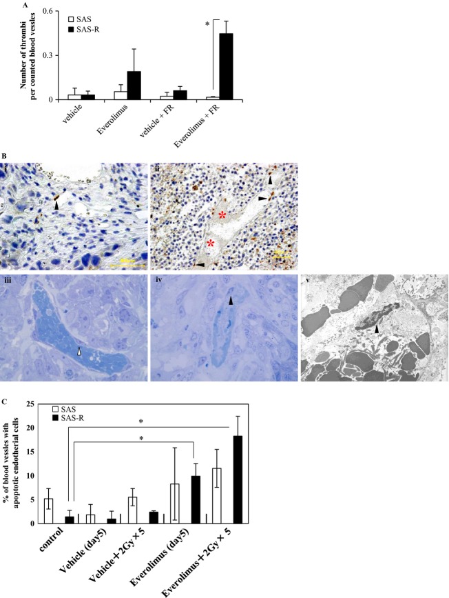
(A) The number of the thrombus per counted blood vessels. The highest density was observed in SAS-R tumors treated with fractionated radiation (FR) and Everolimus. Mean ± SD in triplicate. *P < 0.01. (B-i and ii) Immunohistochemical detection of single-strand DNA-positive cells. Black arrow heads, single-strand DNA-positive endothelial cells. red asterisk, thrombosis. (B-i) SAS tumor 24 h after exposure to 5 days of FR (2 Gy/day) with Everolimus. (B-ii) SAS-R tumor 24 h after exposure to 5 days of FR (2 Gy/day) with Everolimus. Thrombosis was frequently found. (B-iii) Most endothelial cells of blood vessels surrounding the surviving tumor area had no nucleus with condensed chromatin in SAS-R tumors 3 days after starting Everolimus and FR treatment. White arrow head, endothelial cell. (B-iv, v) Some apoptotic tumor cells (e.g., with condensed chromatin) near the central necrosis area were found by vessels with apoptotic endothelial cells containing old thrombosis. Black arrow heads, apoptotic endothelial cells. (iii, iv) Toluidine blue staining. ×1250 magnification. (v) ×4000 magnification. (C) Frequencies of blood vessels with apoptotic endothelial cells. Even Everolimus alone significantly induced endothelial cell apoptosis in SAS-R tumors, however, these endothelial cell apoptosis were remarkably reinforced by Everolimus combining with FR. In contrast to SAS-R tumor tissues, significant induction of apoptotic endothelial cells in SAS tumors was not observed by Everolimus compared with control, even after combining with FR. *P < 0.01. FR, fractionated radiation.

