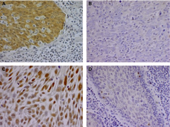
Figure 1.

Representative photographs of p16 and p53 immunohistochemistry staining. A 40× magnification of (A) a p16-positive tumor sample; (B) a p16-negative tumor sample; (C) a sample with 100% of the tumor cells expressing p53; and (D) a sample with 0% of the tumor cells expressing p53.
