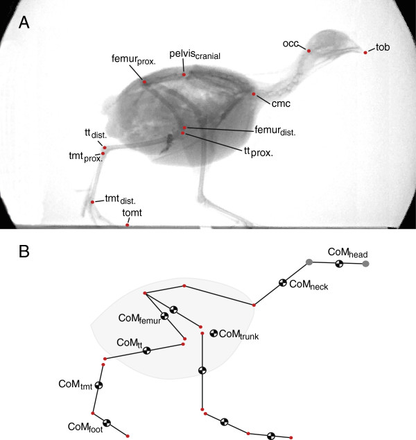
Figure 2.

Kinematic analysis and the kinematic model used for the calculation of the body’s CoM (center of mass). A: Lateral x-ray projection to show the skeletal landmarks digitized. Landmarks were identified and digitized non-automatically. Both hindlimbs were digitized though landmarks for one limb are only shown here. B: Stick figure representation of the kinematic model. The mass and precise CoM position (using a pendulum method) of each segment was determined [23]. The instantaneous position of the body’s CoM was calculated by combining the kinematic data, the mass and inertial properties. Please note that CoMtrunk and the CoM of the whole body are not identical. In simulations only the timing of the head landmarks (grey) were modified with respect to the experimental data (red) of the remaining landmarks. Tob: tip of beak; occ: occiput; cmc: caudal-most cervical vertebra; tt: tibiotarsus; tmt: tarsometatarsus; tomt: tip of middle toe.
