Figure 1.

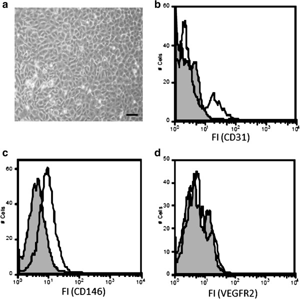
Flow cytometric analyses 2 weeks after isolation and cultivation of rat bone marrow MNC show heterogeneity of the cell population. (a) Cells were grown in specialized endothelial cell growth medium (EGM2 MV) for 2 weeks after isolation and their morphology was examined using light microscopy. The scale bar depicts 100 μm. The cells were further analyzed by Flow Cytometry for expression of endothelial cell-specific surface markers including CD31 (b), CD146 (c) and VEGF-R2 (d). Grey histograms indicate fluorescence signals of negative controls; white histograms indicate fluorescence signals of specific antigens. Results are representative of 4 separate experiments.
