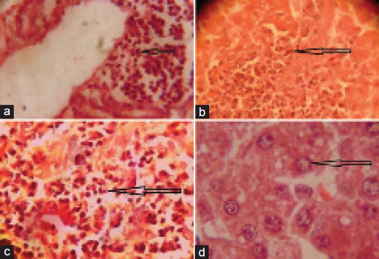
Figure 1.

(a) Microphotograph showing infiltration of mononuclear cells around central vein in liver of rat treated with Sodium arsenite @ 10 mg/kg (×400). (b) Microphotograph showing necrosis, degenerating parenchyma in of rat treated with Sodium arsenite @ 10 mg/kg. (×400). (c) Microphotograph showing the highly necrosed area, degenerating parenchyma and damaged cellular architecture in liver of rat treated with sodium arsenite @ 10 mg/kg. (×400). (d) Microphotograph showing necrosis and vacuolar degeneration in liver of rat treated with sodium arsenite @ 10 mg/kg (×400)
