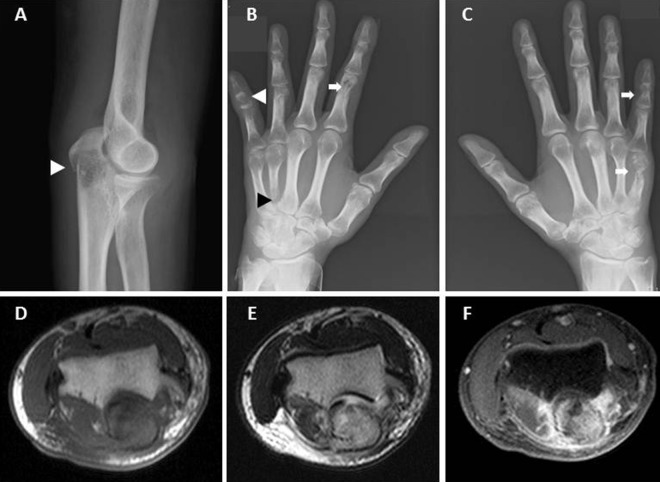
Figure 1.
Lateral radiograph of the right elbow showing the fracture line at the right olecranon (A; white arrowhead) with cystic lesions, suggesting osteolytic changes. Furthermore, radiographs showing multiple osseous erosions (B and C; arrow) with osteolytic lesions, fractures on the middle phalanx of the left digitus minimus manus (B; white arrowhead) and base of the left metacarpal of digitus annularis (B; black arrowhead). Axial T1-weighted imaging (T1WI) demonstrates a low-intensity intramedullary mass measuring 2 cm in the right olecranon (D), appearing hyperintense on axial T2WI (E) and enhancing heterogeneously on fat-suppressed enhanced T1WI (F).

