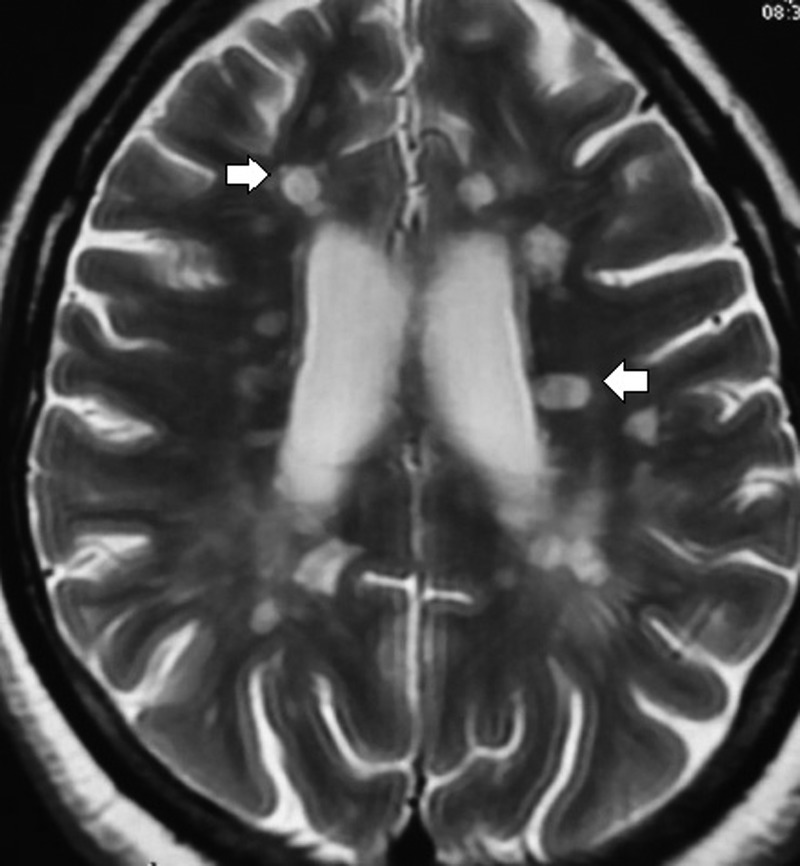
Figure 2.

Axial sequence of T2-weighted image revealed multiple, small discrete, oval, >3 mm hyperintense lesion (demyelinating plaques) in the white matter involving the supratentorial, juxtacortical and periventricular regions and the corpus callosum (white arrows).
