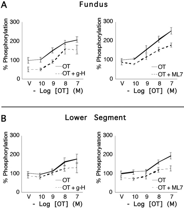
Fig. 4.

Concentrations of pRLC in uterine myocytes from upper and lower uterine segments following stimulation by oxytocin. Primary cultures of myocytes from the uterine fundus and lower uterine segments (n = 5 patients) were treated with increasing concentrations of OT for 20 s and the response measured using a specific antibody for pRLC phosphorylated at S19 in an in-cell western assay. There were significant, concentration-dependent increases that were similar in cells from upper and lower segments. 15-minute pretreatment with the rho-kinase inhibitor (g-H; 1 µM) or the myosin regulatory light chain kinase inhibitor (ML7; 25 µM) caused approximately 20-30% suppression of basal pRLC but the response to OT was maintained.
