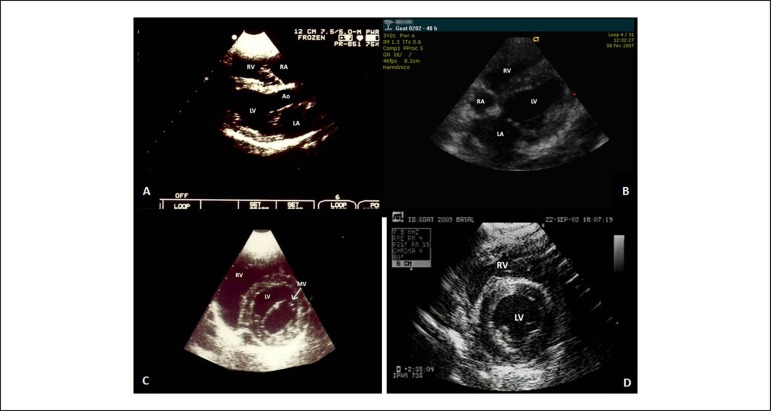
Figure 4.
Echocardiographic views from the right parasternal approach in a dog (A and C) and a goat (C and D). Parasternal long-axis view (A). Left and right ventricular inflow tract long-axis view (B). Parasternal short-axis view at the baseline (C) and at the papillary muscles level (D).MV: mitral valve; RV: right ventricle; LV: left ventricle; Ao: aorta; LA: left atrium; RA: right atrium.

