
Figure 2.
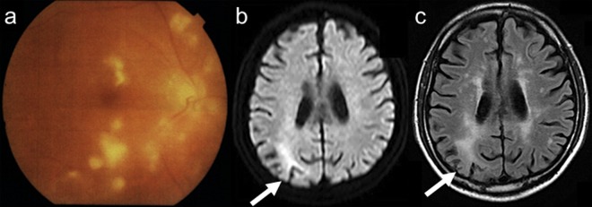
Follow-up of funduscopy examinations (A). Diffuse white-coloured lesions were extended despite antibiotics or corticosteroids. Follow-up images of diffusion-weighted imaging (B) and fluid-attenuated inversion recovery (FLAIR) (C). Restricted diffusion and increased FLAIR signal were seen in the right parietal lobe on admission.