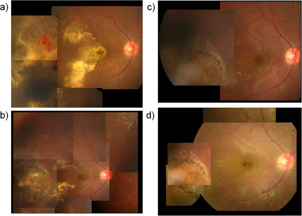
Figure 2.
Case 1 fundus photographs. a) Prior to the injection of intravitreal bevacizumab (IVB). b) Five months after IVB injection. Hard exudates in the posterior pole had gradually resolved. c) Eleven months after IVB injection. Hard exudates in the posterior pole had almost disappeared. d) Eighteen months after IVB injection. Abnormal retinal vessels in the periphery are completely cicatrized and no recurrence is observed.

