Figure 1.
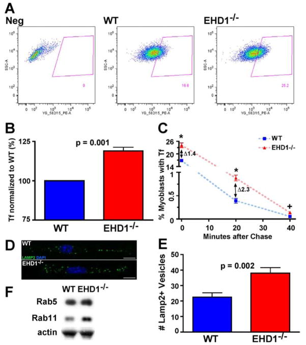
Reduced recycling and lysosome accumulation in EHD1-null myoblasts. A) Wildtype and EHD1-null primary myoblasts were incubated with transferrin conjugated to Alexa-546 for 15 minutes and analyzed for fluorescence intensity by FACS. FACS profiles showed increased transferrin accumulation in EHD1-null myoblasts. B) EHD1-null myoblasts contained approximately 20% more transferrin fluorescence compared to wildtype myoblasts (n=3 replicates, p = 0.001). C) Wildtype and EHD1-null primary myoblasts were pulsed with transferrin conjugated to Alexa-546, chased with unlabelled transferrin for 0, 20, or 40 minutes and then analyzed by FACS for fluorescence intensity. A greater number of EHD1-null myoblasts retained Alexa-546 fluorescence at 0, 20, and 40 minutes consistent with a defect in recycling (* p< 0.03, + p=0.08). D) Lysosomes were identified in wildtype and EHD1-null myoblasts using anti-LAMP2 antibody (green). Nuclei are stained with DAPI (blue). Scale bar 10μm. E) The number of LAMP2-positive vesicles was increased in EHD1-null primary myoblasts (mean number 23 in wildtype myoblasts and 38 in EHD1-null myoblasts, n=13, p=0.002). F) Immunoblot of WT and EHD1-null muscle lysates. WT and EHD1-null muscle show similar Rab5 expression levels. Rab11 expression levels are increased in EHD1-null muscle. Actin is shown as a loading control.

