
Figure 4.
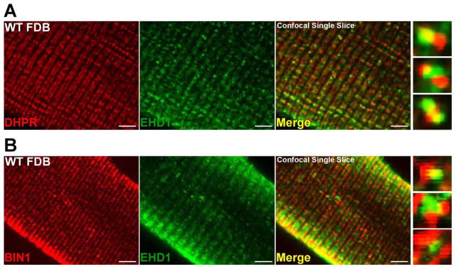
EHD1 localizes to the T-tubule in wildtype muscle. Representative single slice confocal images are shown with high magnification images on the right. A) Myofibers were stained with anti-DHPR (red) and anti-EHD1 (green) antibodies. EHD1 localizes partially with the T-tubule marker, DHPR. B) Additional myofibers were stained with anti-BIN1 (red) and anti-EHD1 (green) antibodies. EHD1 staining partially overlaps with BIN1 at the T-tubule and at the sarcolemma within wildtype myofibers. Scale bar 5μm.