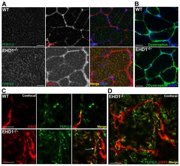
Figure 6.
Abnormal localization of Fer1L5 and caveolin-3 in mature EHD1-null muscle. Mature muscle was stained with anti-Fer1L5 (green) and anti-caveolin-3 (red) antibodies. DAPI is shown in blue. A) Fer1L5 and caveolin-3 internal staining was increased in EHD1-null muscle (bottom row) compared to wildtype controls (top row). Scale bar 30μm. B) Wildtype and EHD1-null muscle were stained with anti-dystrophin (green) and DAPI (blue) to outline the myofibers, confirming the presence of a uniform sarcolemma in both samples. Scale bar 30μm. C) Fer1L5 and caveolin-3 colocalized in EHD1-null muscle at the membrane in vesicular patterns and within the muscle fiber on tubule structures (bottom row; merge, arrow). Scale bar 1μm. D) Low magnification image of an EHD1-null myofiber with excessive internal Fer1L5 and caveolin-3 tubules. Scale bar 10μm.

