
Figure 7.
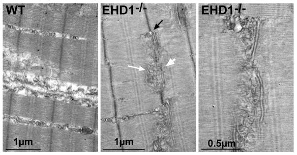
Enlarged sarcoplasmic reticulum and T-tubular structures in the absence of EHD1. Representative electron microscopy images of swollen sarcoplasmic reticulum (black arrow), elongated T-tubules (white arrowhead), and vesicular structures (white arrow) juxtapose the T-tubules in EHD1-null quadriceps muscle. This was never seen in wildtype controls. A magnified view of the EHD1-null disrupted sarco-tubular system is shown in the right panel.