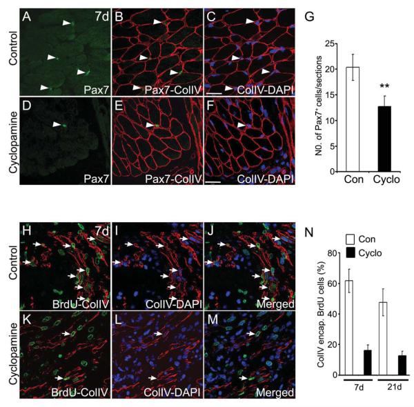
Fig. 5. Pax7+ cell populations and regenerating fibers are reduced upon Hh inhibition.
(A-F) Immunohistochemical analysis for Pax7+ cells (green) and collagen type IV [colIV (red)] basal lamina from control (A-C) and cyclopamine (D-F) treated regenerating tissue at 7d. (C, F) shows nuclei [DAPI (blue)] merged with colIV+ lamina. Arrowheads indicate the position of the Pax7+ cells. (G) Quantification of the Pax7+ cells in the regenerating limbs from control and cyclopamine treated tissues. Note the reduced number of Pax7+ cells in the cyclopamine treated tissue (n=4; **, p<0.005). (H-M) Longitudinal sections of regenerating limb tissue from control (H-J) and cyclopamine (K M) treated animals at 7d, stained with anti-BrdU (green) and anti-colIV (red) antibody. Nuclei are stained with DAPI (blue). (J, M) show the combined images of BrdU (green), colIV (red) and DAPI (blue) channels. Regenerating fibers are indicated by arrows. (N) Quantification of the percentage of colIV-encapsulated-BrdU+ cells (intralaminar) at 7d and 21d regenerating tissue. Error bars indicate s.e.m. Scale bar: 50 μm.

