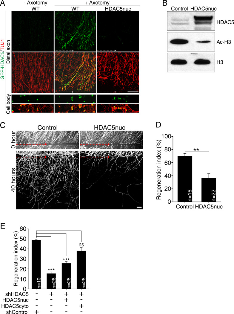
Figure 5. Expression of a nuclear HDAC5 mutant decreases the level of histone H3 acetylation and impairs axon regeneration in vitro.
(A) DRG spot cultures overexpressing GFP-HDAC5 (WT) or GFP-HDAC5nuc mutant were axotomized and stained with TUJ1. White dotted arrow, axotomy line. Scale, 100µm. (B) DRG neurons expressing GFP (control) or GFP-HDAC5nuc were analyzed by western blot. (C) DRG spot cultures expressing GFP only (control) or GFP together with GFP-HDAC5nuc were axotomized and fixed 40 hours after axotomy. Axons were visualized by GFP live imaging. Red dotted arrow indicates the axotomy site. Scale, 100µm. (D) In vitro regeneration index calculated from images in (C) (**p<0.01). (E) In vitro regeneration index calculated as in (D) in control (shControl), HDAC5 knock-down (shHDAC5), HDAC5 knock-down plus GFP-HDAC5nuc and HDAC5 knock-down plus GFP-HDAC5cyto (***p<0.001; ns, not significant). Data are represented as mean±SEM. See also Figure S2.

