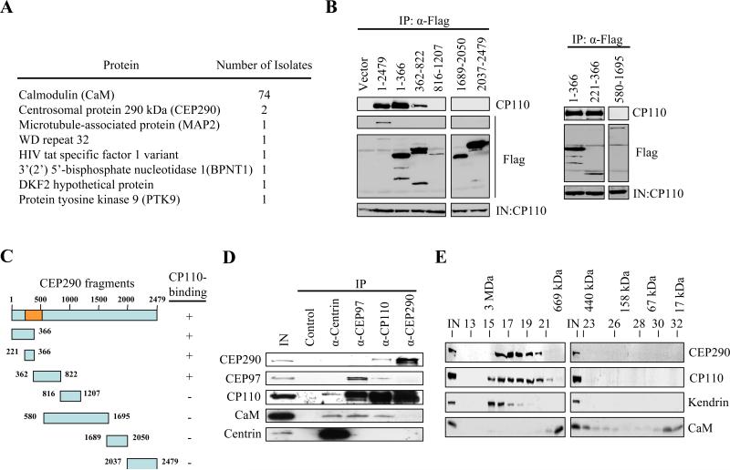
Figure 1.
CP110 interacts with CEP290 in vivo. (A) The number of times that each annotated protein was identified in the yeast two-hybrid screen is shown. (B) The indicated fragments of Flag-tagged CEP290 were expressed in 293T cells and immunoprecipitated from lysates. Flag-CEP290 fusion proteins and CP110 were detected after western blotting the resulting immunoprecipitates. Input CP110 was detected in lysates from each transfection (IN). (C) Summary of CEP290 truncation mutants and the results of in vivo binding experiments. The orange box denotes the CP110-binding domain based on the yeast two-hybrid screen. (D) Western blotting of endogenous CEP290, CEP97, CP110, CaM, and centrin after immunoprecipitation with anti-Flag (control), anti-centrin, anti-CEP97, anti-CP110, or anti-CEP290 antibodies from 293T cell extracts. IN represents input. (E) Cell extract was chromatographed on a Superose 6 gel filtration column, and the resulting fractions were blotted with antibodies against CP110, CEP290, kendrin, or CaM. Estimated molecular weights are indicated at the top of the panel.

